
Development
Finding the cells that put our brain to sleep
September 18, 2026 Development Source: Ars Technica
Share this article



“Going after these cells was really a high-risk project because the likelihood of seeing anything with 0.1 percent of neurons in the cortex is really low,” Batista-Brito said. “I wrote a bunch of grants on these projects that were always rejected because it was too high-risk.”
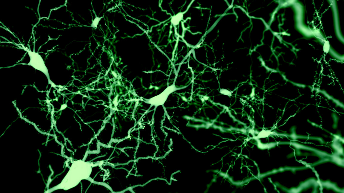
Her first look at the anatomy of Sst-Chodl neurons proved the risk worth taking. “At first, I saw two or three cell bodies in the whole brain,” Batista-Brito told Ars. “Despite that, there was massive, massive arborization all over the visual cortex, like I never saw with any other neuron.”
That “massive arborization” is a tree-like branching that connects neurons to other parts of the brain, making them part of a wider communication network. It was also very unusual, as inhibitory neurons are almost always local. They receive inputs from different, sometimes distant regions in the brain and exert control over their immediate surroundings—their own small patch.
“Sst-Chodl neurons are kind of the opposite,” Batista-Brito said. “They are receiving inputs that are quite precise, but then they are broadcasting that information everywhere.”
A single Sst-Chodl cell branches out to the entire visual cortex, then sends axons to the areas of the brain responsible for touch, hearing, spatial memory, navigation, and voluntary movements.
To see when the cells fire, the team imaged them with while tracking the mice’s pupil size, muscle tone, facial movement, running, and cortical electrical activity at the same time. Ninety-five out of 111 imaged cells lit up during slow-wave sleep and quiet, motionless wakefulness, and fell silent during running and REM sleep.
During slow-wave sleep, the cortex alternates between UP states of vigorous firing and DOWN states of near-silence; the Sst-Chodl neurons broke this pattern. “Their activity is even higher at the termination of the UP state,” Terral said. “Once all the other neurons start to decrease their activity, those neurons start to increase it even more.”
They also skip the rebound that other cells show coming out of a DOWN state. “Those cells behave totally differently to any other cells we can measure around that transition,” Terral said.
To really understand what they do, the team made the SSt-Chodl neurons fire on command using optogenetics, which involves inserting the gene for a light-activated ion channel that can stimulate the nerve cells to fire.
Activating the cells across the cortex of freely moving mice increased slow-wave and REM sleep, cut the time taken to fall asleep, and sent the animals into their nests during the day. Then the team tried it during the dark phase, when nocturnal mice are mostly awake.
“We could have them sleep more during the time that they are awake than they usually sleep during the day,” Batista-Brito said. “I never thought this experiment was going to work. We are manipulating one percent of inhibitory neurons with local injections, so we are hitting only a fraction of those cells. And we could see an effect that was really quite striking.”
That effect, Batista-Brito argues, points toward the idea that these neurons might be the cortex’s sensors of sleep pressure, something first proposed by Thomas Kilduff, the director of the SRI International’s Center for Neuroscience and co-author of the study.
Sleep, Batista-Brito explains comes in two flavors: the circadian sort that follows the light and homeostatic sleep, the accumulated fatigue that eventually makes us sleep whether we want to or not. Kilduff, Batista-Brito said, showed that after sleep deprivation, these are the most active cells in the cortex.
One question Batist-Brito and her colleagues still don’t have the answer to, though, is what activates these cells.
“One caveat of our work is that it was done in the visual cortex,” Batista-Brito said.
The findings about the coordinating role of the Sst-Chodl neurons, the team claims, should generalize to different brain regions, but the actual wiring may not. Batista-Brito’s lab is now repeating the anatomy part of their work in the prefrontal cortex. Their prediction is that there we’ll see inputs from other areas, such as the hypothalamus and the thalamus, and that will be what triggers the network for sleep specifically.
The cells are conserved from salamanders to humans, which is why the paper argues they could be an entry point into the sleep disruption running through so many psychiatric illnesses.
The team is now focused on three remaining questions: what activates these cells, whether they actually sense sleep pressure, and why they drive delta power.
Nature, 2026. DOI: 10.1038/s41586-026-10876-y