
Development
Toddler's tragic death from brain-destroying amoeba offers lessons for doctors
August 13, 2026 Development Source: Ars Technica
Share this article



One of the biggest missteps was a misdiagnosis early in his case. Often in these rare amoeba cases, the early symptoms are so vague that diagnoses are delayed. But in the boy’s case, doctors jumped to a diagnosis—the wrong one—based on an unusual presentation. When his parents brought him to an emergency room, his symptoms were vomiting, fatigue, lethargy, and weakness. A computed tomography (CT) scan showed he had a hemorrhage on his brain, requiring a shunt.
More imaging of his vasculature showed that some of his arteries were blocked or narrowed. This has not been reported with amoeba infections before. Doctors ended up diagnosing him with childhood Takayasu arteritis, a type of vasculitis. It’s a rare, systemic inflammatory condition that causes damage to large and medium arteries in the body. It wasn’t a perfect diagnosis, but it was the condition that fit best with what they could see. They treated the boy with immunosuppressive drugs, and his symptoms improved in the hospital. After about two weeks in the hospital, he went home.
However, a week later, he was back in an emergency department with vomiting. Doctors sent him home after tests found his shunt was working properly, suspecting it was likely a side effect of one of the medications for his inflammatory condition. But his parents brought him back two days later, this time with fever, lethargy, a headache, and trouble swallowing. He was re-admitted to the hospital, and his condition began to deteriorate. At this point, it had been 25 days since his first hospital visit. The next day, he needed to be intubated. Two days after that, day 28 of his illness, he became unresponsive, his eyes rolled upward with pinpoint pupils, and his muscles became floppy.
Doctors scrambled to figure out what was going on, running more tests. Imaging of his brain suggested his injuries were worsening. They upped his dose of immunosuppressive therapy, thinking it was due to his vasculitis. But it wasn’t helping as it did before, and they took him off it as they began to worry he could have a fungal infection.
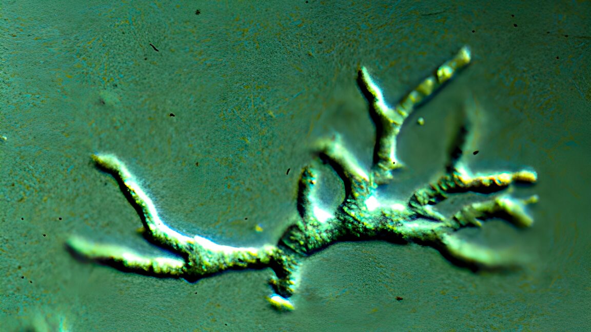
Thirty days after his initial hospital visit, doctors performed a brain biopsy to try to find the cause of his condition. They also turned to metagenomic next-generation sequencing (mNGS) on samples from his cerebrospinal fluid, a test that picks up and reads any genetic material in the fluid to determine what might be in there. Both tests finally picked up the rare amoeba, Balamuthia mandrillaris. In the brain tissue, doctors could see the amoebas, and their genetic signature was in his brain, too. Once the amoeba was identified, doctors began a large regimen of antimicrobial and antiparasitic drugs. But by then, catastrophic damage had been done.
To keep this from happening again, his doctors recommend that before immunosuppressive treatment is started or escalated, doctors should do diagnostic testing on cerebrospinal fluid for infectious microbes. The genetic testing of the cerebrospinal fluid was positive, suggesting this could be a relatively noninvasive way to identify the amoeba in future cases.
Early detection of a B. mandrillaris can make a difference of life and death for children. In 2010, doctors in Kentucky reported a case in a 2-year-old boy who survived. His infection was identified 10 days after he was brought to a hospital. Although he had a significant neurologic injury, he made improvements after he was discharged from the hospital. At discharge, he was unable to follow commands, communicate, or walk. After 22 months, he was able to hold himself upright, smile socially, follow simple commands, and was working on talking.
In these cases—and almost all others—it’s unclear how or why the patients became infected. B. mandrillaris lives in soil and water around the world, though cases are mostly seen in warm places. People are thought to get infected by inhaling the amoeba from windblown dust or by getting contaminated soil or water in a wound. Experts do not know why some people become infected and others don’t. People with compromised immune systems are at higher risk, but healthy people, like the boy in the new case report, can also become infected.
Common initial symptoms of B. mandrillaris begin with fever, headache, vomiting, nausea, and tiredness. In some cases, people develop telltale skin lesions, particularly on the face. Neurologic symptoms can develop days, weeks, or months later, including changes in mental status, confusion, seizures, paralysis, and difficulty walking and/or talking.